| |
Chapter Ten | Delgado Index | Chapter Twelve
Jose Delgado's "Physical Control of the Mind"
Methodology for Direct Communication with the Brain
The depth of the central nervous system can be reached very easily through the natural windows of sensory receptors. Stimuli such as patterns of light travel fast front the eye's retina through optic pathways to the visual cortex located in the occipital lobe. Would it be possible to explore the local activity of cortical neurons during the process of symbolic perception? Could we evoke similar sensations by direct stimulation of specific neurons? Can we reach the mind of an individual without using the normal ports of sensory entry? Can we direct the functions of the brain artificially? These and similar problems have attracted the interest of many investigators, but the brain is well protected by layers of membranes, spinal fluid, bone, and teguments, a formidable shield which for a long time has kept the secrets of mental functions away from scientific curiosity.
Implantation of Electrodes in Animals
Starting in the last century, many investigators have explored the brain, first in animals and recently in human patients as part of diagnosis and therapy. In these studies it was necessary to open both skin and skiill, and because the procedure was painful it was mandatory to use anesthesia. It blocked pain perception, but it also inhibited some of the most important
functions of the nervous system. Emotions, consciousness, and free behavior were certainly absent under heavy sedation, and for many years scientists directed their attention to sleeping subjects and overlooked the complexity of awake brains. Textbooks of cerebral physiology were concerned with pathways, connections, reflexes, posture, and movement; mental functions and behavior were considered to belong to a different discipline.
The methodological breakthrough which made it possible to study the brain of behaving animals came in the 1930s when W.R. Hess (106) devised a procedure to implant very fine wires within the brain of anesthetized cats. After the effects of anesthesia had disappeared, the relatively free and normal animal could be electrically stimulated by connecting long leads to the terminals of the implanted electrodes. This procedure was refined in the early 1950s (47, 49) by reducing the size of the electrodes while increasing the number of intracerebral contacts and using aseptic precautions during implantation. Surgical accuracy in reaching chosen cerebral targets was also improved by means of micromanipulators and a precise system of anatomical coordinates which made it possible to reach similar structures in different subjects. Using biologically inert materials such as gold, platinum, or stainless steel wires insulated with teflon allows the electrodes to be left inside of the brain indefinitely. A diagram of the cerebral implantation of one assembly of seven contacts is shown in Figure I and the X ray of the head of a monkey after implantation is seen in Figure 2. Through a small opening in the skull, the shaft is introduced down to a predetermined depth and is secured with dental cement at the point where it passes through the skull. Then the upper portion of the shaft is bent over the bone surface and secured again a short distance away, and the terminal socket is exteriorized on the head. Each contact of the socket corresponds to a determined point in the depth of the brain which is accessible merely by plugging in a connector, a procedure as simple as connecting any electrical appliance to a wall outlet. This technique has been used for ESB in thousands of animals in
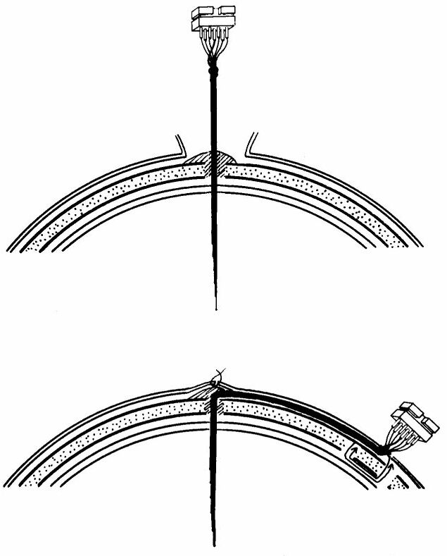
Diagramatic representation of an electrode assembly implanted within the brain and anchored to the skull. The depth of the brain is thus accessible simply by plugging in a connector (52).
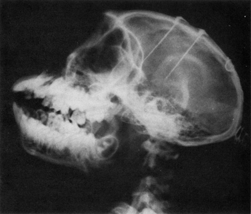
X rays of a monkey's head showing two assemblies of electrodes implanted in the frontal lobes and in the thalamus (49).
many laboratories around the world, and there is ample experience of its efficiency, accuracy, and safety, resolving the initial skepticism that introduction of wires into the brain would be technically difficult, dangerous for the subject, and grossly disruptive of normal functions. It is true that implantation of electrodes destroys neurons along the path of penetration, breaking capillary vessels and later producing a local reaction involving the formation of a thin fibrotic capsule along the implantation tract. It has been proven, however, that local hemorrhage is neglible and that because of the well-known functional redundancy of neural tissue with abundance of duplication in its circuits, the destruction of a relatively small group of neurons
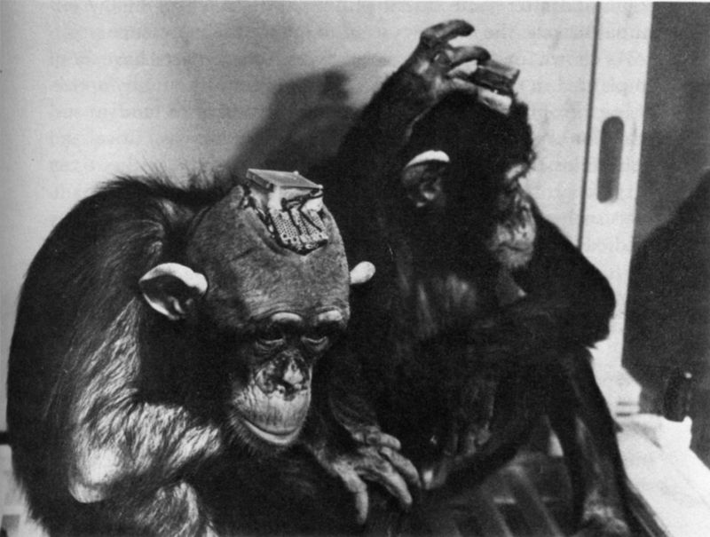
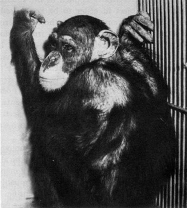
Chimpanzees Paddy (left) and Carlos, each with too intracerebral electrodes and boxes for instrumentation. in spite of this massive implantation, no detectable behavioral deficits have been found, and the animals are still alive and in excellent health two years after surgery.
does not produce any detectable deficit. The thin reactive encapsulation is electrically conductive and is not an obstacle to stimulation or recording. Beyond this 0.1-0.2 millimeter capsule, the brain appears histologically normal. judged by the absence of abnormal electrical activity, the reliability of effects evoked by ESB, and the consistency of thresholds of excitability through months of experimentation, the electrodes seem to be well tolerated. Some of our monkeys have had electrodes in their heads for more than four years. The anchorage is very
solid, and after some initial pulling and scratching of the terminal sockets, the monkeys seem to ignore their presence.
As shown in Figure 3, as many as 100 contacts have been implanted in the brain of some chimpanzees without any noticeable neurophysiological or behavioral disturbance, and in several monkeys, contacts have been placed in areas as critical and delicate as the respiratory centers of the medulla without any surgical problems. Electrodes have been used in laboratory animals such as rats, cats, and monkeys, and also in less frequently studied species including crickets, roosters, dolphins, and brave bulls.
Electrodes in the Human Brain
Our present knowledge of the central nervous system is based principally on investigations in animals. Experience has shown that many questions about implantation in humans, such as biological tolerance of electrodes by the neural tissue, can be successfully answered in cats or in lower species. Some of the electrochemical events of neural conduction can be analyzed just as adequately in squids as in mammals, and for certain studies of memory, the octopus has proven an excellent subject. The rat has been - and still is - the animal preferred by experimental psychologists because it is a small and inexpensive mainmal which can be used in large quantities to provide behavioral results suitable for statistical evaluation. The limited behavioral repertory of lower animals, however, cannot be compared with the complex activities of monkeys and apes. These species, being closer relatives of man, are more appropriate subjects for the neurophysiological study of intelligent behavior, and when we want to investigate the highest psychological functions of the brain which involve verbal communication, there is no possible substitute for man himself.
The human brain, like any other part of the body, may suffer traumatic accidents, rumors, or illnesses, and it has often been necessary to explore the affected areas in order to identify structtires, assess abnormality of the tissues, test excitability, and
learn the location of important functions which should not be disrupted during surgical procedures. Conscious participation of the patient was required in some of these explorations, for example, to ascertain whether the aura of epileptic attacks could be triggered by electrical stimulation of a specific cortical point@ thus providing information about the possible source of epileptic discharges which could be removed by surgery. For this kind of investigation the brain was exposed under local anesthesia, presenting an exceptional opportunity to study behavioral and psychological responses evoked by ESB in fully awake subjects. The most extensive work in this area has been carried out by Penfield and his associates in Montreal (174), and a considerable number of similar studies have been performed by other neurosurgeons as well (2, 8, 97, 124, 163, 215).
Exploration of the exposed brain has, however, some obvious limitations. It has to be brief to avoid prolongation of surgery; electrodes are usually held in place by hand, causing variability in the applied mechanical pressure; the exposed brain is subject to possible thermal, mechanical, and chemical trauma; the cortical areas are identified only by visual inspection; and the physical and psychological stress of the patient undergoing operation introduces factors difficult to control. Most of these handicaps can be avoided with the use of implanted electrodes, and given the experience of aiiinial experimentation it was natural that sorile investigators should contemplate the application of this methodology to patients for diagnostic and therapelitic purposes (19, 59, 98). Neurosurgeons had already proved that the central nervous system is not as delicate as most people believe, and during therapeutic surgery parts of the cerebral tisstie have been cut, frozen, cauterized, or ablated with negligible adverse effects on the patients. Exploratory introduction of needles into the cerebral ventricles is a well-known and relatively safe clinical procedure, and since electrodes are smaller in diameter than these needles, their introduction into the brain should be even less traumatic. Experience has confirmed the safety and usefulness of long-term implantation of electrodes in man, and the procedurehas been used in specialized medical centersaround
the world to help thousands of patients suffering from epilepsy, involuntary movements, intractable pain, anxiety neurosis, and other cerebral disturbances. In general several assemblies of fine electrodes with a total of twenty to forty contacts are placed on the surface and/or in the depth of the brain, with the terminal connectors exteriorized through the scalp and protected by a small head bandage (see Figure 4). In some cases the electrodes have remained for nearly two years with excellent tolerance.
Leaving wires inside of a thinking brain may appear unpleasant or dangerous, but actually the many patients who have undergone this experience have not been concerned about the fact of being wired, nor have they felt any discomfort due to the presence of conductors in their heads. Some women have shown their feminine adaptability to circumstances by wearing attractive hats or wigs to conceal their electrical headgear, and many people have been able to enjoy a normal life as outpatients, returning to the clinic periodically for examination and stimulation. In a few cases in which contacts were located in pleasurable areas, patients have had the opportunity to stimulate their own brains by pressing the button of a portable instrument, and this procedure is reported to have therapeutic benefits.
Chronically implanted electrodes enable careful diagnostic explorations to be performed without time limit, and repeated electrical excitations or well-conttolled coagulations can be graded according to the reactions of the patient. As a bonus, important information about psychophysiological correlations, providing direct knowledge about the cerebral basis of human behavior, is being acquired. In our studies (60, 109, 150), an interview situation was selected as the method most likely to offer a continuous supply of verbal and behavioral data. While the electrical activity of eight pairs of cerebral points was being recorded, we taped about one hour of conversation between therapist and patient. Notes of the observable behavior were also taken, During the interview, electrical stimulations of the brain were applied for 5 seconds with intervals of three or more minutes, and each significant point was explored several times.
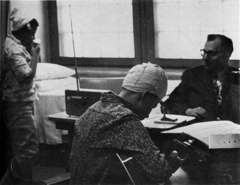
Two girls who were suffering from epileptic seizures and behavioral disturbanccs requiring implantation of electrodes in the brain for diagnostic and therapeutic purposes. Under the cap, each patient wean a "stimoceiver," used to stimulate the brain by radio and to send electrical signals of brain activity by telemetry while the patients are completely free within the hospital ward (60). One example of electrical recordings is shown in Figure 17.
Two-way Radio Comunication with the Brain
Electronic technology has reached a high level of sophistication, and two-way radio communication with automobiles, airplanes, and outer space vehicles is commonplace today. The notable lag in development of similar instrumentation for
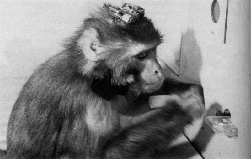
A monkey instrumented with 28 implanted electrodes, a two-channel telemetric unit on top of the head, and a three-channel radio stimulator around the neck. The animal has learned to press a lever to obtain fund. With this methodology, brain functions can be explored by remote control without disturbing the behavior under observation.
communciation with the depth of the brain reflects the already mentioned unbalanced evolution of our technological civilization, which seems more interested in accumulating power than in understanding and influencing the basic mechanisms of the human mind.
This gap is now being filled, and as Figures 4 and 5 show, it is already possible to equip animals or human beings with minute instruments called "stimoceivers" for radio transmission and reception of electrical messages to and from the brain in completely unrestrained subjects. Microminiaturization of the instrument's electronic components permits control of all param-
eters of excitation for radio stimulation of three different points within the brain and also telemetric recording of three channels of intracerebral electrical activity. In animals, the stimoceiver may be anchored to the skull, and different members of a colony can be studied without disturbing their spontaneous relations within a group. Behavior such as aggression can be evoked or inhibited. In patients, the stimoceiver may be strapped to the head bandage, permitting electrical stimulation and monitoring of intracerebral activity without disturbing spontaneous activities.
Stimoceivers offers geat promise in the investigation, diagnosis, and therapy of cerebral disturbances in man. Preliminary information about use in patients with temporal lobe seizures (see Figure 4) has demonstrated the following advantages over other methods of intracerebral exploration (60): (1) The patient is instrumented simply by plugging the stimoceiver to the head sockets. (2) There is no disturbance of the spontaneous individual or social behavior of the patient. (3) The subject is under continuous medical supervision, and stimulations and recordings may be made day and night. (4) Studies are carried out during spontaneous social interactions in the hospital environment without introducing factors of anxiety or stress. (5) The brain in severely disturbed patients may be explored without confinement to a recording room. (6) As connecting wires are not necessary there is no risk of dislodgment of electrodes during abnormal behavior. (7) Therapeutic programed stimulation of the brain can be prolonged for any necessary amount of time.
It is reasonable to speculate that in the near future the stimoceiver may provide the essential link from man to computer to man, with a reciprocal feedback between neurons and instruments which represents a new orientation for the medical control of neurophysiological functions. For example, it is conceivable that the localized abnormal electrical activity which announces the imminence of an epileptic attack could be picked tip by implanted electrodes, telemetered to a distant instrument
room, tape-recorded, and analyzed by a computer capable of recognizing abnormal electrical patterns. identification of the specific electrical disturbance could trigger the emission of radio signals to activate the patient's stimoceiver and apply an electrical stimulation to a determined inhibitory area of the brain, thus blocking the onset of the convulsive episode.
This speculation is supported by the following experiments completed in June, 1969, in collaboration with Drs. Johnston, Wallace, and Bradley. Chimpanzee Paddy (Figure 3), while free in her cage, was equipped with a stimoceiver to telemeter the brain activity of her right and left amygdaloid nuclei to an adjacent room, where these waves were received, tape-recorded, and automatically analyzed by an on-line analog computer. This instrument was instructed to recognize a specific pattern of waves, a burst of spindles, which was normally present in both amygdaloid nuclei for about one second several times per minutes The computer was also instructed to activate a stimulator, and each time the spindles appeared, radio signals were sent back to Paddy's brain to stimulate a point in her reticular formation known to have negative reinforcing properties. In this way electrical stimulation of one cerebral structure was contingent on the production of a specific EEG pattern by another area of the brain, and the whole process of identification of information and command of action was decided by the on-ine computer.
Results showed that about two hours after the brain-to-computer-to brain feedback was established, spindling activity of the amygdaloid nucleus was reduced to 50 per cent; and six days later, with daily two-hour periods of feedback, spindles were drastically reduced to only 1 per cent of normal occurrence, and the Chimpanzee was quieter, less attentive, and less motivated during behavioral testing, although able to perform olfactory and visual tasks without errors.
The computer was then disconnected, and two weeks later the EEG and Paddy's behavior returned to normal. The experiment was repeated several times with similar results, sup-
porting the conclusions that direct communication can be established between brain and computer, circunivening normal sensory organs, and also that automatic learning is possible by feeding signals directly into specific neuronal structures without conscious participation.
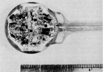
One of the limiting factors in these studies was the existence of wires leading from the brain to the stimoceiver outside of the scalp. The wires represented a possible portal of entry for infection and could be a hindrance to hair grooming in spite of their small size. It would obviously be far more desirable to employ minute instruments which could be implanted completely beneath the skin. For this purpose we have developed in our laboratory a small three-channel stimulator which can be placed subcutaneously and which has terminal leads to be implanted within the liraiii (Figure 6). The instrument is solid state, has no batteries, and can work indefinitely. Necessary electrical energy, remote control of parameters of stimulation, and choice of channels are provided by transdermal coupling, using a small coil which is activated by frequency-modulated radio signals. In February, 1969, an experiment was begun in monkey Nona and in chimpanzee Suzi who were equipped with subcutaneous stimulators to activate their brains from time to time for the rest of their lives. Terminal contacts were located in motor pathways in order to evoke flexion of the contralateral leg, an effect simple enough to be observed and quantified without difficulty. Study of Nona and Suzi and preliminary investigations in other animals have demonstrated that subcutaneous instrutiientation is efficient, reliable, and well tolerated. Behavioral responses were consistent, and local motor excitability was not modified by repeated experimentation. Thus the technical problems of stimulating any desired area of the brain for as long as necessary in the absence of conductors passing through the skin have been solved, therapeutic and scientific possibilities have been multiplied, and the comfort of subjects has been considerably increased.
The next technical step will be to combine transdermal
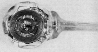
Figure 6
Both sides of a three-channel transdermal stimulator. This instrument has no batteries, is activated by radio, and can be used for life, so that the brain can be stimulated indefinitely. Chimpanzee Suzi (right) has two units (six channels) implanted on her back underneath the skin.
stimulation of the brain with transdermal telemetry of EEG. In this case the stimoceiver will not be outside the skin as it was in Paddy (Figure 3), nor will it be limited to only transdermal stimulation (Figure 6) as in Nona and Suzi: the whole instrument will be totally subcutaneous. The technology for nonsensory communication between brains and computers through the intact skin is already at our fingertips, and its consequences are difficult to predict. In the past the progress of civilization has tremendously magnified the power of our senses, muscles, and skills. Now we are adding a new dimension: the direct interface between brains and machines. Although true, this statement is perhaps too spectacular and it requires cautious clarification. Our present knowledge regarding the coding of information, mechanisms of perception, and neuronal bases of behavior is so elemental that it is highly improbable that electrical correlates of thoughts or emotions could be picked
up, transmitted, and electrically applied to the suitable structure of a different subject in order to be recognized and to trigger related thoughts or emotions. It is, however, already possible to induce a large variety of responses, from motor effects to emotional reactions and intellectual manifestations, by direct electrical stimulation of the brain. Also, several investigators have learned to identify patterns of electrical activity (which a computer could also recognize) localized in specific areas of the brain and related to determined phenomena such as perception of smells or visual perception of edges and movements. We are advancing rapidly in the pattern recognition of electrical correlates of behavior and in the methodology for two-way radio communication between brain and computers.
Fears have been expressed that this new technologies with it the threat of possible unwanted and unethical remote control of the cerebral activities of man by other men, but as will be discussed later, this danger is quite improbable and is outweighed by the expected clinical and scientific benefits. Electronic knowledge and microminiaturization have progressed so much that the limits appear biological rather than technological. Our greatest need is for more experimental information about the neuronal mechanisms related to behavioral and mental processes, and research in unrestricted subjects promises to reveal new understanding of normal minds and more efficient therapy of disturbed brains.